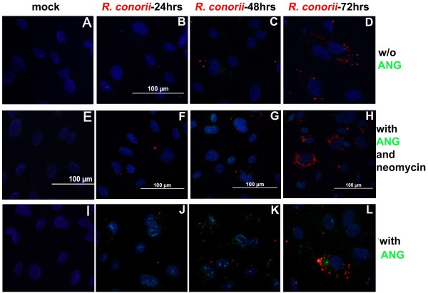
Figure 3.
SFG rickettsial infection initiated compartmentalized translocation of exogenous rANG in human primary endothelial cells. Dual immunofluorescence staining of SFG rickettsiae (red) and ANG (green) in human umbilical vein endothelial cells (HUVECs) using a dual wave lengths filter system revealed that there was no significant detectable endogenous ANG in endothelial cells (images A-D). R. conorii infection triggered compartmentalized translocation of exogenous rANG at different times p.i. (images J-L). Neomycin reduced cellular internalization of exogenous rANG in SFG rickettsiae-infected endothelial cells (images F-H). Nuclei of HUVECs are counter-stained with DAPI (blue).

