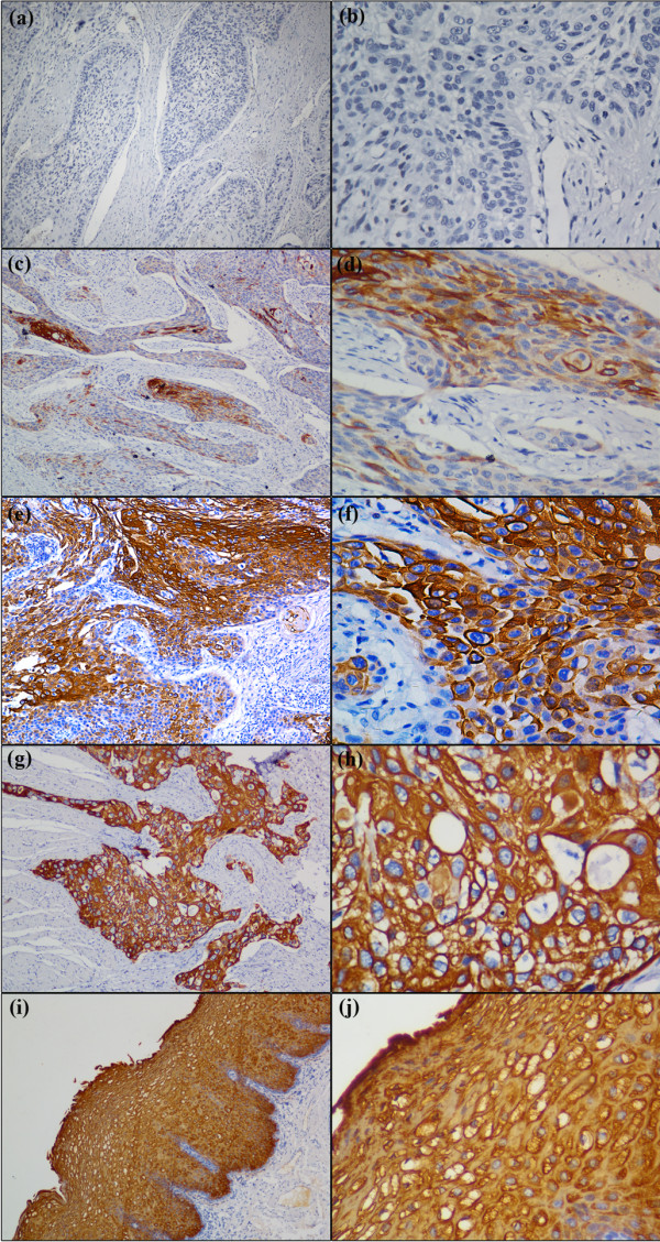
Figure 1.
Immunohistochemical staining of esophageal squamous cell carcinoma (ESCC) and peritumoral normal esophageal mucosae with anti-Cystatin SN. Low expression of Cystatin SN were detected in ESCC tissues (a, b and c, d), and the IRS grades of (a, b) and (c, d) belong to absent(−) and weak(1+), respectively, in which (a, c) original magnification is × 100, and (b, d) original magnification is × 400, respectively; High expression of Cystatin SN was detected in ESCC tissues (e, f and g, h) and peritumoral normal esophageal mucosae( i, j ), and the IRS grades of (e, f) and (g, h and i, j) belong to moderate (2+) and strong (3+), respectively, in which (e, g, i) original magnification is × 100, and (f, h, j) original magnification is × 400, respectively.

