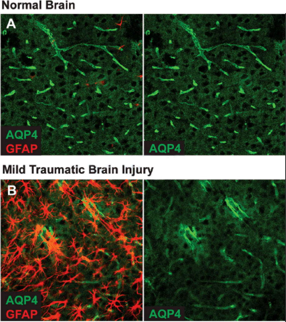
Figure 2. Changes in AQP4 localization after diffuse injury.
(A) Immunofluorescent double-labeling demonstrates that in the healthy young mouse brain, AQP4 expression is highly localized to perivascular astrocytic endfeet surrounding to entirety of the cerebral microvasculature. (B) 7 days after mild traumatic brain injury, widespread reactive astrogliosis (GFAP-immunoreactivity) is observed throughout the ipsilateral cortex. In regions of reactive astrogliosis, AQP4 localization is severely perturbed, exhibiting a loss of polarization to the endfoot process and increased somal labeling. Similar expression patters are observed after diffuse microinfarction (manuscript under review).

