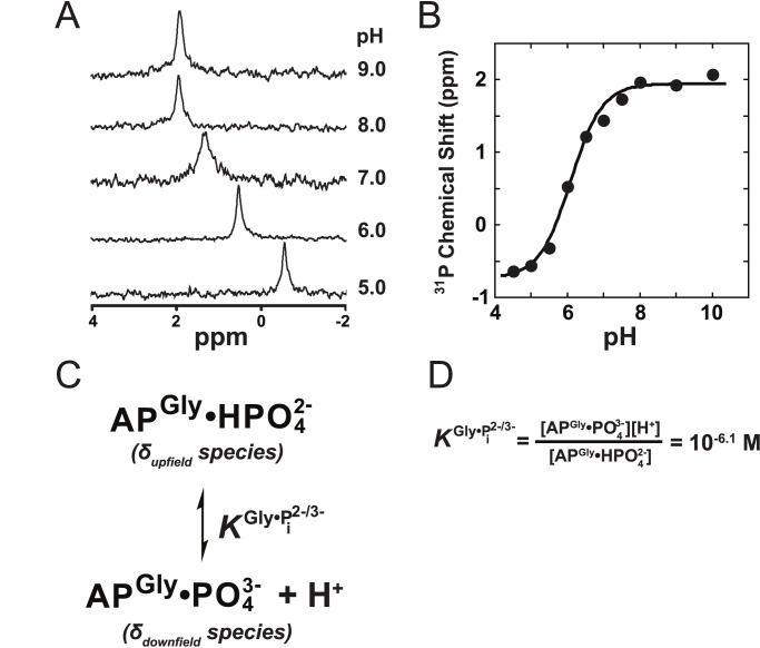
Figure 6. The pH-dependence of the 31P chemical shift of Pi bound to S102G/R166S AP.
(A) 31P NMR spectra of Pi bound to S102G/R166S AP at various pH values. See Materials and Methods for conditions. (B) The chemical shift of Pi-bound to S102G/R166S AP versus the solution pH. A nonlinear least-squares fit to an equation (see Materials and Methods) derived from the binding model in (C) yields δupfield and δdownfield values of −0.74 and 1.94 ppm, respectively, and a value for  of 10−6.1 M as defined in (D).

