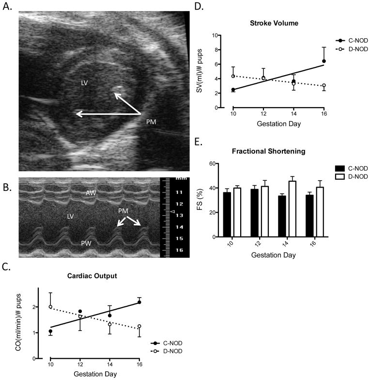
FIG. 1.
Maternal Cardiac Analyses. (A) An example of a B-Mode image of parasternal short axis view of the heart from a gd16 d-NOD. M-Mode sample volume was placed over the middle of the chamber, slightly off the papillary muscle. (B) M-Mode cine loop of parasternal short axis of the heart, also from a gd16 d-NOD. Measurements were performed excluding papillary muscle, seen as caps above the posterior wall in systole. (C) Cardiac output normalized by viable implant site number. Increasing CO is seen over gestation in c-NOD (p=0.025), but not d-NOD mice. Effect of gestation (analyzed via regression slopes) differed between groups (p=0.024). (D) Stroke volume normalized for viable implant site number. Increasing SV is seen over gestation in c-NODs (p=0.035). Again, no increase in SV is observed over gestation in d-NODs. Effect of gestation was different between groups (p=0.030). (E) Fractional shortening. FS was increased in d-NOD compared to c-NODs across the study (p=0.0265), however no significance was reached at any specific gd. No effect of gestation is seen on FS in either d-NOD or c-NOD dams. PM = Papillary muscle, LV = Left ventricle, AW = Anterior left ventricular wall, PW= Posterior left ventricular wall. Solid line and closed circles = c-NODs, dashed lines and open circles = d-NODs. Significantly different slopes upon regression analysis indicated differing effects of gestation between groups, slopes by linear regression considered significantly non-zero at p<0.05.

