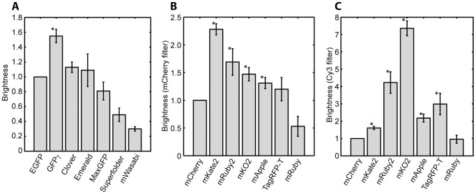
Figure 2. Brightness of green and red fluorescent proteins.
Yeast expressing fusions of each of the optimized fluorescent proteins to the TDH3 protein were imaged, and the mean fluorescence of each strain was calculated. Data from each day was normalized to EGFP (for green proteins) or mCherry (red proteins) to compensate for day-to-day fluctuations in lamp brightness and detection efficiency. The measurement was repeated on at least three days and the mean and standard error for each strain is plotted. * indicates a protein significantly brighter than EGFP or mCherry as determined by a one-sided t-test with 5% significance threshold. A. Green fluorescent proteins. B. Red fluorescent proteins imaged with an mCherry filter set. C. Red fluorescent proteins imaged with a Cy3 filter set.

