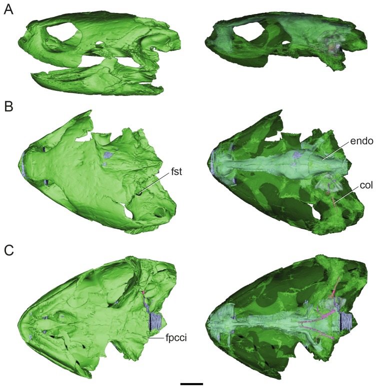
Figure 1. Volume-rendered CT-based reconstruction of the skull of the extinct turtle Plesiochelysetalloni (MH 435).
In the images of the left side the bone is rendered opaque, whereas in the right side the bone is rendered semitransparent to show the endocranial cavity. The bone is shown in green, the cranial endocast and inner ear in light blue, the internal carotid arteries in pink, and the columella in fuchsia. Skull in left lateral (A) dorsal (B) and ventral (C) views. Abbreviations: col, columella auris; endo, cranial endocast; fpcci, foramen posterius canalis caroticus internus; fst, foramen staedio-temporale. Scale bar equals 10 mm.

