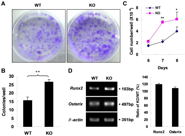
Figure 6. APN deficiency enhances MSC proliferation and osteoblastic differentiation.
(A) Crystal violet staining shows an increased number of CFU-F in MSCs from APN KO mice. (B) The graphs indicate the number of positive colonies/well by crystal violet staining. (C) MSC proliferation assay by cytometry shows increased proliferation of APN KO MSCs. *indicates p<0.05, **indicates p<0.01. (D) RT-PCR shows that up-regulation of two transcriptional factors Runx2 and Osterix in MSCs from APN KO mice. A representative result of three independent experiments is shown.

