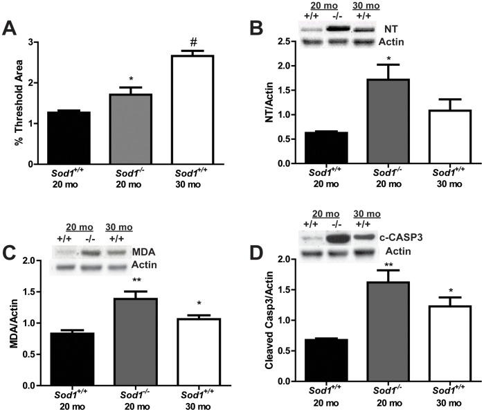
Figure 2. Assessment of oxidative damage in the motor neuron micro-environment.
Oxidative damage was assessed in the spinal cord of in Sod1 mice by A) quantitatively assessing the autofluorescence of lipofuscin, and by western immunoblotting of B) nitrated protein (NT) and C) oxidative lipid degradation (MDA). The Sod1 +/+ mice at 20 mo, Sod1 −/− mice at 20 mo, and Sod1 +/+ mice at 30 mo are represented by white, black, and gray bars, respectively. D) Apoptosis was assessed in the motor neurons of Sod1 mice by cleaved caspase-3 western immunoblotting. Densitometry revealed an increase in cleaved caspase-3 in the in Sod1 −/− mice at 20 mo compared to the Sod1 +/+ mice at 20 mo. *p<0.05, **p<0.01; n≥4 for all groups.

