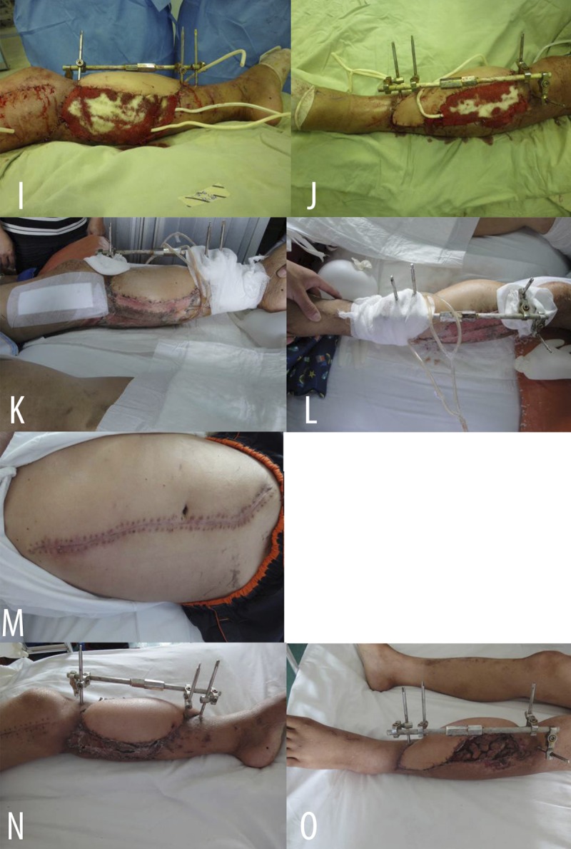
Figure 2I–O.

(I, J) Covered remnant wounds around the flap with skin grafts; covered recipient site with VSD (I – medial view; J – lateral view). (K, L) 5d after operation (K – medial view; L – anterolateral view). (M) 2 weeks after operation. Donor site of thoraco-umbilical flap healed. (N, O) 2 weeks after operation. Flap and skin grafts successfully grew, infection was well controlled and subcircular wound achieved primary repair (N – medial view; O – lateral view).
