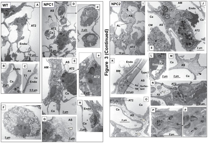
Figure 3. Morphology of wild type and mutant mice lungs by electron microscopy.
Wild type lung (A–C). A. Overview of section of lung with alveolar type II cell (AT2) and endothelial cell (Endo). B. Capillary (Ca). C. Respiratory membrane with type I cell (T1) and endothelial cell (Endo). AS, alveolar space. NPC1 mutant lung (D–H). D. Overview of lung with leukocytes in the capillary and excess surfactant in alveolar space. AL, alveolar lipidosis. d. Enlargement of area in D showing vacuolar leukocyte (L). Similar leukocytes were seen in NPC2 mutant lung. E. Alveolar type II cell (AT2) with a foamy alveolar macrophage (AM) in close proximity in the alveolar space. e. Enlargement of area in E showing type II cell-macrophage contact. *Indicates vacuolar inclusions in endothelial cell. F. Alveolar macrophage with lipid-like material and vacuolar inclusions. G. Type II cells with excess surfactant (white arrowhead). H. Endothelial cell with vesicular inclusions (*). NPC2 mutant lung (I–P). I. Overview of lung with surfactant completely filling the alveolar space characteristic of alveolar lipidosis. J. AT2 with an alveolar macrophage containing multivesicular whirls and a foamy circulating macrophage (CM). K. Respiratory membrane of endothelial cell, basement membrane (BM) and type I cell and demonstrating large amounts of surfactant as tubular myelin (TM) and aggregate (Ag) structures. L. Endothelial cell with vesicular structures. M. Alveolar space with black arrowhead indicating proteinaceous material. Similar material was seen in NPC1 mutant lung. N. Large aggregate structure with tightly packed phospholipid-type whirls (gray arrowheads) or string-like structures (white arrowhead) in alveolar space. O. Surfactant vesicles (white arrowheads) filling the alveolar space. P. Type II cell with inset (p) showing autophagosome-like structures (ap) in enlargement.

