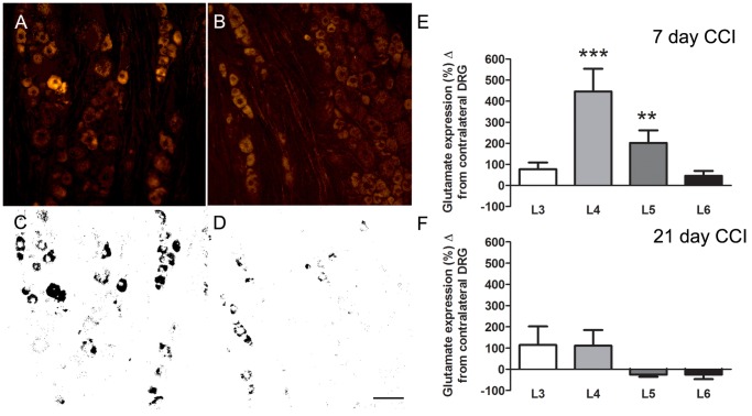
Figure 7. Representative images from an L4 DRG immunostained for glutamate following CCI of the sciatic nerve.
Increased glutamate immunolabeling is seen in the DRG ipsilateral to the sciatic CCI (A standard, C thresholded image) compared to the contralateral side (B standard, and D thresholded image). E, F shows the % difference in glutamate immuno-expression between the ipsi and contralateral L3 - L6 DRGs at 7 (E) and 21 (F) day post-CCI-SN animals. Scale bar = 50 µm. Data expressed as mean ± SEM. **, P<0.01; ***P<0.001 ipsilateral vs. contralateral.

