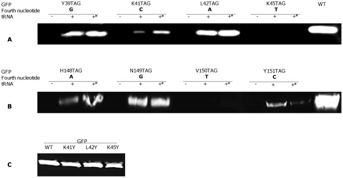
Figure 3. Cell-free expression of WT GFP and tyrosine-incorporating mutant GFP, as visualized by Western blot.
(A and B) Co-translational incorporation of tyrosine at different positions in response to the amber stop codon was achieved by adding purified MjTyrRS (200 µg/mL) and two types of suppressor tRNA (480 µg/mL) to the reaction mixture (tRNA denotes synthetic MjtRNACUA, *– tRNACUA Opt). (C) Western blot visualization of the expression level of GFP WT and tyrosine-substituted proteins.

