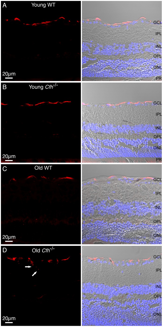
Figure 5. GFAP expression in astrocytic glial cells in Cfh −/− and wild-type mice.
12 µm sections from young (A and B) and one year (C and D) wild-type and Cfh −/− fixed mouse eyed were stained for GFAP (red) and nuclei (blue). Sections were imaged using confocal and differential interference contrast microscopy. Arrows indicate astrocytic processes extending into IPL. GCL, ganglion cell layer; IPL, inner plexiform layer; INL, inner nuclear layer; OPL, outer plexiform layer; ONL, outer nuclear layer; PR, photoreceptors. Images are representative of three independent experiments. Scale bar = 20 µm.

