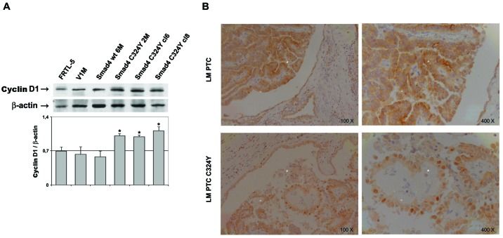
Figure 2.
Expression of cyclin D1. (A) Whole protein lysates (60 μg/lane) from FRTL-5, V1M and Smad4 stable clones (wt 6M, C324Y 2M, C324Y cl6, C324Y cl8) were analyzed by western blotting using an antibody against cyclin D1. Densitometric evaluation of the cyclin D1 signals was performed normalizing to the levels of β-actin [* indicates a statistical significance (Student’s t-test, P<0.05) of Smad4 C324Y clones vs. control cells]. (B) Immunohistochemistry, using antibody against cyclin D1, was performed in lymph node metastasis of PTC from which derives the C324Y mutation of Smad4 and in a group of 3 lymph nodal metastases of PTC without mutation. Original magnification, ×100 (panels on the left) and ×400 (panels on the right).

