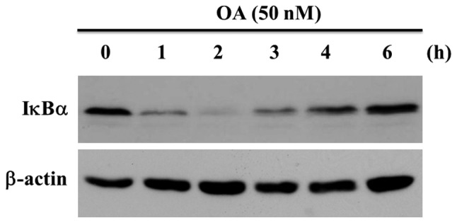
Figure 2.

Degradation of IκBα in MG63 cells treated with OA. New protein synthesis in MG63 cells was inhibited by 100 ng/ml of CHX for 30 min. The cells were treated with 50 nM OA for various time courses as indicated (upper panel). The samples from the OA-treated cells were analyzed by western blotting with anti-IκBα antibody. The loading control done with β-actin is shown in the lower panel.
