
Figure 4.
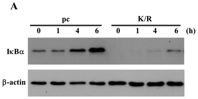

Expression of IκBα in pc and PKR-K/R cells. (A) The pc and PKR-K/R cells were treated for various time periods with 50 nM OA. The expression of IκBα was analyzed by western blotting. Loading controls were done with anti-β-actin antibody. (B) The expressions of IκBα mRNA in MG63 and PKR-K/R cells were analyzed by real-time PCR. Significant differences from the control cultures are indicated by asterisks; *P<0.05; **P<0.01; WT, MG63 cells; KR, PKR-K/R cells.