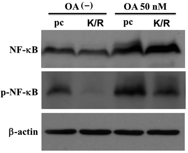
Figure 5.

Western blot analysis of NF-κB in OA-treated cells. The pc and PKR-K/R cells were treated with 50 nM OA for 6 h and the cell lysates were prepared from each type of culture. Twelve microgram of each sample was separated on a 10% of SDS-PAGE and transferred to PVDF membranes. Each membrane was then incubated with anti-p65NF-κB (NF-κB) and anti-phospho-Ser536 NF-κB (p-NF-κB) antibodies. The membrane antibody was stripped off and re-incubated with anti-β-actin antibody as a loading control.
