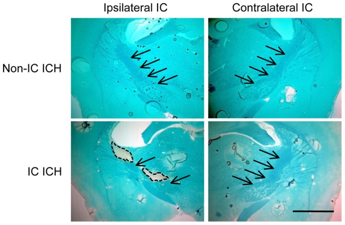
Figure 5. Axonal damage associated with invasion of hematoma into IC.

Shown are representative images of Luxol fast blue staining of coronal sections (−1.4 mm relative to bregma) obtained from a non-IC ICH mouse and an IC ICH mouse at 4 weeks after induction of ICH. Arrows indicate IC, and broken lines indicate areas damaged by hematoma. Scale bar = 1 mm.
