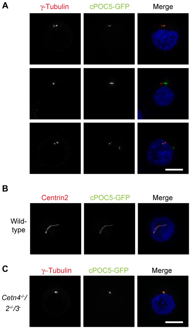
Figure 5. cPOC5 overexpression induces linear structures.

A. Immunofluorescence micrograph shows examples of the structures induced by transient transfection of DT40 cells with a cPOC5-GFP expression construct. GFP is shown in the green channel, centrosomes were identified by γ-tubulin staining (red) and DNA was visualised with DAPI (blue). Micrographs are representative of results obtained in at least 2 separate experiments. Scale bar, 5 µm.
B. Centrin colocalisation with cPOC5-GFP structures was observed by microscopy for centrin2 (red), with cells otherwise being transfected, fixed and stained as for A.
C. Centrosomal localisation of cPOC5-GFP was observed in centrin-deficient cells. Cells were transfected, fixed and stained as for A.
