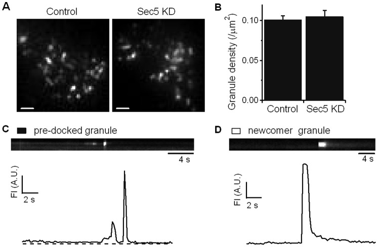
Figure 3. Sec5 depletion does not affect recruitment of insulin granules to dock onto the plasma membrane.
(A) TIRF imaging of exocytosis of Control and Sec5 KD INS-1 832/13 cells infected with Ad-IAPP-mCherry. Scale bar 5 µm. (B) The graph shows a comparison of averaged granule densities from control and Sec5 KD INS-1 cells before stimulation (n = 10 cells for each). (C–D) Kymographs and the corresponding time-lapse fluorescence intensity (FI) curves indicate different fusion modes. (C) Fusion event of a pre-dock insulin granule; (D) fusion event of a newcomer granule that did not undergo a docking step on the plasma membrane before proceeding to exocytosis. A.U., arbitrary units.

