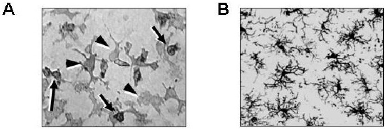
Fig. 1.
Microglia in culture and in the brain. (A) Microglia cultured from neonatal rat brains were stained with anti-CD11b antibodies. Microglia show two different morphologies: round (arrows) and irregular-shaped (arrowheads). (B) Brain sections obtained from 8-week-old rats were stained with anti-Iba-1 antibodies. Highly ramified microglia are detectable in the cortex.

