
Figure 1.
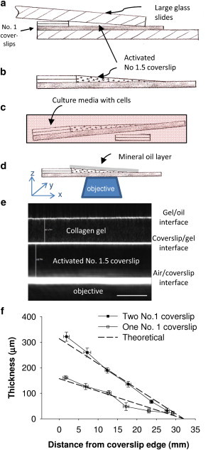
(a) Schematic of the experimental setup for creating the sloped-gel sample. The collagen or fibrin gel adheres to the activated No. 1.5 coverslip and is formed into the sloped shape by the upper large glass slide that is weighted with a 50 g object. (b) Schematic of the finished sample ready for cell seeding. (c) Samples are placed in a 100 mm petri dish and the right side is propped up with two No. 1 coverslips to ensure a level seeding surface. (d) Sample setup for imaging with mineral oil to minimize reflection at the edge of the gel. (e) Confocal reflectance image taken in cross-section mode shows the No. 1.5 coverslip between the bottom and middle white lines, and protein gel fibers with speckled pattern between the top two lines (10× dry objective; scale bar = 100 μm). The change in height (∼4 μm) cannot be readily seen over the field of view (∼400 μm) due to the gradual slope of the gel. (f) Validation of the slopes of fibrin gels created in the system using either one or two No. 1 coverslips.