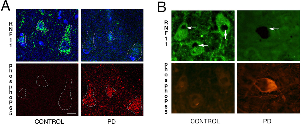
Fig 3. Reduced RNF11 is associated activated NF-κB in PD.
Cortical (A) and nigral (B) sections from brains of control (left panel) and PD (right panel) cases were double labeled for RNF11 and phospho-p65, a marker for activated NF-κB pathway. (A) Confocal images in the top panel demonstrate RNF11 immunoreactivity (green) and bottom panel demonstrates phospho-p65 immunoreactivity (red) in cingulate cortex. Nuclei were stained with Hoechst 333258 (blue). (B) Photomicrographs of nigral sections from brains of control (left panel) and PD (right panel) patients demonstrating immunoreactivity for RNF11 (green, top panel) and phosph-p65 (red, bottom panel). Nigral dopaminergic neurons are identified by dense neuromelanin pigmentation (arrow). These representative images demonstrate that in PD brain reduced RNF11 immunoreactivity is associated with increased phospho-p65 immunoreactivity. The ‘gain’ was adjusted to avoid over-saturation while taking fluorescent images. Scale bar of (A) − 10µm and (B) − 20µm.

