
Figure 3.
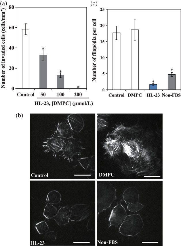
Inhibitory effects of HL-23 on the invasion of LM8 cells in vitro. (a) In vitro invasion assay. Invaded cell number of LM8 cells after the treatment of HL-23 for 24 h. Error bars indicate SE for three individual experiments. *P < 0.05 to control (5% glucose solution). (b) Micrographs of LM8 cells stained with rhodamine–phalloidin after the treatment of HL-23 for 3 h by TIRF microscopy. Scale bars: 20 μm. (c) Number of filopodia on the surface of LM8 cells. Error bars indicate SE for 8–14 individual experiments. *P < 0.05 to control. Control: 5% glucose solution, DMPC liposomes: [DMPC] = 50 μmol/L, HL-23: [DMPC] = 50 μmol/L, [C12(EO)23] = 5.6 μmol/L, non-FBS: LM8 cells were incubated in the serum-starved medium for 24 h. HL, hybrid liposomes; LM8, murine osteosarcoma; SE, standard error; TIRF, total internal reflection fluorescence; DMPC, l-α-dimyristoylphosphatidylcholine; C12(EO)23, polyoxyethylene(23) dodecyl ether; FBS, fetal bovine serum.