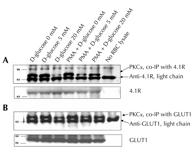
Figure 4. Co-immunoprecipitations of PKCα with GLUT1 and 4.1R.

RBC lysates were exposed to protein A-Sepharose coupled to anti-GLUT1 or anti-4.1R. Immunoprecipitates were analyzed by SDS-PAGE followed by Western blot, as described under "Materials and methods". A representative result is presented in the figure. Similar results were obtained in two additional experiments. Data of 4.1R, GLUT1, and light chain of correspondent antibodies were used as controls to verify equivalent amounts of protein throughout the lanes. In the control 'no lysate' study, protein A-Sepharose coupled to an appropriate antibody was exposed to a RBC-free sample. Molecular mass markers are indicated in kDa.
