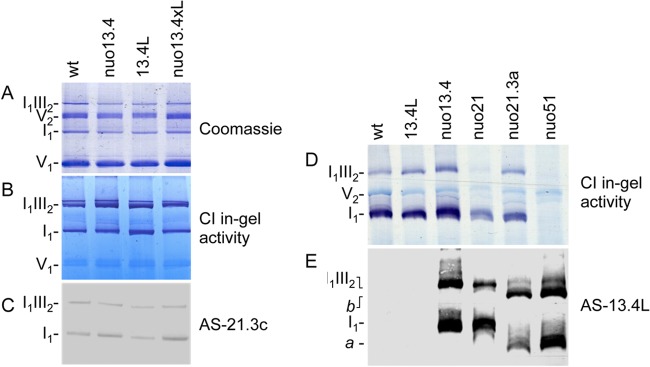
Fig 4.

Effects of 13.4L disruption in respiratory chain organization. Mitochondrial proteins were separated in a 4-to-13% BN PAGE (A, B, and C) or 4-to-9% BN PAGE (D and E). The gels were stained with Coomassie blue (A), stained for NADH dehydrogenase activity (B and D), or analyzed by Western blotting with antibodies against complex I subunit 21.3c kDa (C) and the 13.4L protein (E). a and b are high-molecular-weight structures recognized by AS-13.4L.
