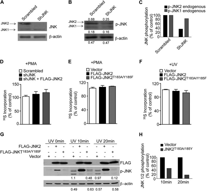
Fig 5.
JNK2 is not required for global translation. (A, B, and C) Levels of endogenous JNK1 and JNK2 expression (A) and activity (B) were monitored by Western blotting in control cells (Scrambled) and cells depleted of endogenous shJNK2 by shRNA (shJNK). Densitometric values are appended to the respective bands. (C) The densitometric values indicated in panel B were normalized to β-actin and are expressed as a percentage of the control (Scrambled). shJNK reduced JNK2 activity by 60%, while only a slight reduction of JNK1 was detected. (D, E, and F) Equal 35S incorporation at the 0-h time point shows that there was no major effect on protein synthesis in the indicated cell lines. Values are represented as mean values ± the SEM (n = 3). (G) HEK293T cells were transfected with FLAG-JNK2 WT or FLAG-JNK2T183A/Y185F, irradiated with UV, and JNK phosphorylation was monitored by Western blotting at the indicated time points. Portions (30 μg) of the corresponding protein extracts were loaded onto SDS-PAGE gels. β-Actin served as a loading control. Densitometric values corresponding to phosphorylation of endogenous JNKs in cells transfected with empty vector or FLAG-JNK2T183A/Y185F are indicated below the respective lanes and are summarized in panel H. JNK phosphorylation was reduced by 50 and 90% after 10 and 20 min, respectively.

