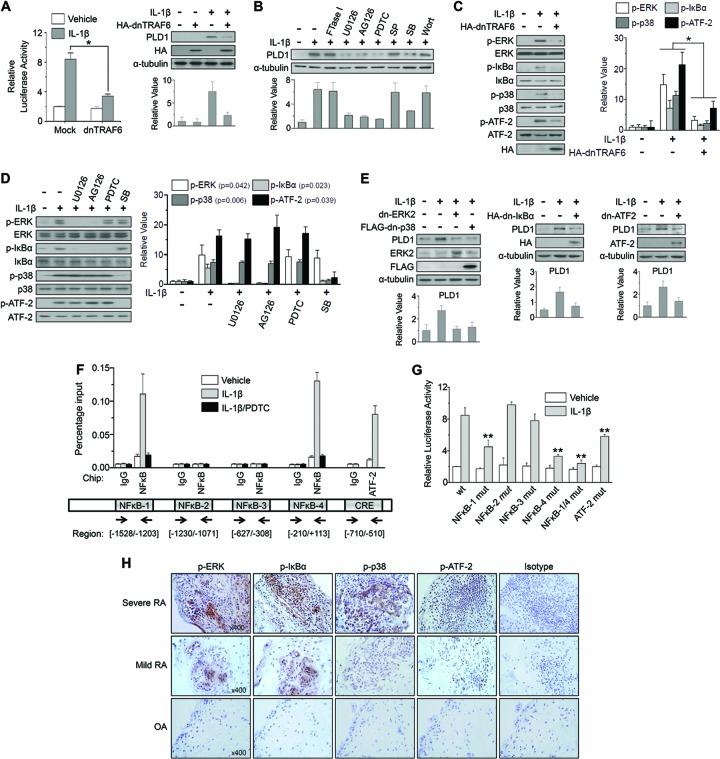
Fig 2.
IL-1β-induced PLD1 expression is mediated by the TRAF6/ERK/NF-κB and TRAF6/p38/ATF-2 signaling pathways. (A) RAFLS were cotransfected with pGL4-PLD1 and dn-TRAF6 and treated with IL-1β (10 ng/ml) for 36 h. A luciferase activity assay was performed (left). PLD1 expression was analyzed by Western blotting (right). (B) RAFLS were pretreated with FTase I, U0126, AG126, PDTC, SP600125 (SP), SB203580 (SB), and wortmannin (Wort) for 30 min and treated with IL-1β for 36 h, after which the lysates were analyzed by Western blotting with an anti-PLD Ab. (C) RAFLS were transfected with dn-TRAF6 and treated with IL-1β. HA, hemagglutinin. (D) RAFLS were pretreated with the indicated inhibitors and treated with IL-1β. (E) RAFLS were transfected with the indicated dominant negative constructs and then stimulated with IL-1β for 36 h. (C to E) Lysates were immunoblotted with the indicated Abs. (A to E) Relative PLD1 protein levels were quantitated by densitometer analysis. The data shown are representative of three independent experiments. (F) RAFLS were pretreated with PDTC for 30 min and then treated with or without IL-1β for 12 h. A ChIP assay was performed with preimmune IgG, anti-NF-κB, or anti-ATF-2 Ab, and the product was analyzed by qPCR. Arrows indicate the positions of the primers used in the ChIP experiment. (G) RAFLS were cotransfected with WT pGL4-PLD1, one or two NF-κB or ATF-2 binding site mutant forms (mut) of pGL4-PLD1 and then treated with or without IL-1β. A luciferase activity assay was then performed. (H) IHC staining of the indicated proteins in synovium from mild or severe RA and OA patients (×400). *, P < 0.01; **, P < 0.05. The data presented are the means ± SDs of four independent experiments.

