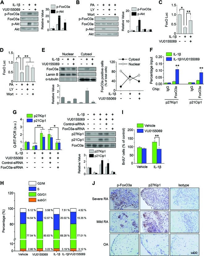
Fig 6.
Inhibition of PLD1 suppresses IL-1β-induced phosphorylation of FoxO3a and enhances cell cycle arrest via transactivation of FoxO3a. (A) RAFLS were pretreated with VU0155069 (10 μM) and stimulated with IL-1β for 30 min. (B) RAFLS were pretreated with a PI3K inhibitor (wortmannin [Wort], LY294002 [LY]) and stimulated with PA (50 μM) for 30 min. (A and B) Lysates were immunoblotted with the indicated Abs. The relative levels of the indicated proteins were quantitated by densitometer analysis. The data shown are representative of four independent experiments. (C and D) RAFLS were transfected with FoxO-Luc and treated with the indicated drugs, and a luciferase activity assay was performed. (E) RAFLS were pretreated with VU0155069 and treated with IL-1β for 30 min. Lysate was fractionated into the cytosol and nucleus and then immunoblotted with the indicated Abs (left). Relative FoxO3a protein levels were quantitated by densitometer analysis. The data shown are representative of four independent experiments. The FoxO3a-positive cells identified in different fields by IHC were quantitated (right). (F) RAFLS were pretreated with VU0155069 and treated with IL-1β for 12 h, and a ChIP assay was performed with the indicated Abs. (G) RAFLS were transfected with or without siRNA specific to FoxO3a and subjected to real-time qPCR (left) and immunoblotting (right). a.u., arbitrary units. The relative levels of the indicated proteins were quantitated by densitometer analysis. The data shown are representative of four independent experiments. RAFLS were treated with or without VU0155069 and IL-1β for 36 h and then analyzed by FACS (H) and BrdU incorporation assay (I). (J) IHC staining of p-FoxO3a and p27Kip1 in the synovium from mild/severe RA and OA patients. *, P < 0.01; **, P < 0.05. The data presented are the means ± SDs of four independent experiments.

