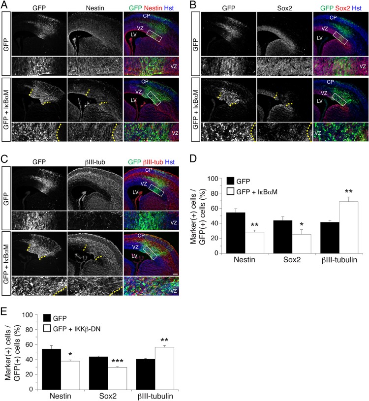
Fig 2.
NF-κB signaling is important to prevent premature neuronal differentiation in the developing neocortex in vivo. (A to C) Double-label immunofluorescence analysis of GFP and either nestin (A), Sox2 (B), or βIII-tubulin (βIII-tub) (C) expression 48 h after in utero electroporation of E13.5 mouse embryos with either a bicistronic plasmid expressing both IκBαM and GFP or the empty vector expressing GFP alone. Bottom rows, high-magnification views of the areas indicated by rectangles in the right-hand panels. Where shown (when IκBαM was used), dotted lines roughly demarcate the GFP+ electroporated area in the VZ. Hst, Hoechst. Scale bar, 100 μm. (D) Quantification of the fraction of GFP+ cells coexpressing nestin, Sox2, or βIII-tubulin. Results are shown as the means ± standard errors of the means (SEM) (*, P < 0.05; **, P < 0.01; n = 5 electroporated embryos per condition; t test). (E) Quantification of the fraction of GFP+ cells coexpressing nestin, Sox2, or βIII-tubulin 48 h after in utero electroporation of E13.5 embryos with either a bicistronic plasmid expressing both IKKβ-DN and GFP or the empty vector expressing GFP alone. Results are shown as the means ± SEM (***, P < 0.001; n = 3 electroporated embryos per condition; t test).

