Fig 6.

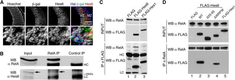
Hes6 is expressed in neocortical progenitor cells in which NF-κB is activated and forms complexes with NF-κB subunit RelA. (A) Double-label immunofluorescence analysis of β-Gal and Hes6 expression in the neocortex of E13.5 NF-κBLacZ embryos. The bottom row depicts high-magnification views of β-Gal and Hes6 coexpression in VZ cells. MZ, mantle zone. Scale bars, 200 μm or 25 μm, as indicated. (B) Nuclear extracts from dissected cortices from E14.5 CD1 mouse embryos were subjected to immunoprecipitation (IP) with anti-RelA or control antibody. Immunoprecipitates were analyzed with each input lysate by Western blotting (WB) with anti-RelA (top) or anti-Hes6 (bottom) antibody. The arrow points to the position of the Hes6 immunoreactive band. HC, immunoglobulin heavy chain; LC, immunoglobulin light chain. (C) Protein extracts from HEK293 cells transfected with empty FLAG vector (FLAG), FLAG-tagged Hes6 (FLAG-Hes6), or FLAG-tagged Hes1 (FLAG-Hes1) were subjected to immunoprecipitation with anti-FLAG antibody. Immunoprecipitates (bottom two panels) were analyzed together with each input lysate (top two panels) by Western blotting with anti-RelA or anti-FLAG antibodies. (D) Protein extracts from HEK293 cells transfected with empty FLAG vector (FLAG), FLAG-tagged wild-type Hes6 (WT), FLAG-Hes6 lacking most of helix 2 (i.e., amino acids 55 to 95) (Δ55-95), FLAG-Hes6 lacking the WRPW motif required for Gro/TLE binding (ΔWRPW), or FLAG-Hes1 were subjected to immunoprecipitation with anti-RelA antibody. Immunoprecipitates (bottom two panels) were analyzed together with each input lysate (top two panels) by Western blotting with anti-RelA or anti-FLAG antibody.
