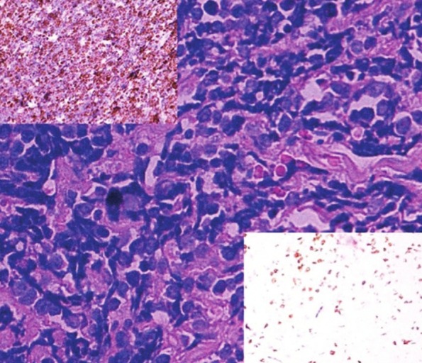
Figure 2.

Sheets of atypical lymphoid cells with hyperchromatic nuclei and scant cytoplasm (H and E, × 400). The upper insert shows increased number of CD 20 positive cells (IHC × 100). Only a few scattered cells in the background are positive for CD 3 in the lower insert (IHC × 100). This staining pattern is diagnostic of NHL – Diffuse large B-cell type
