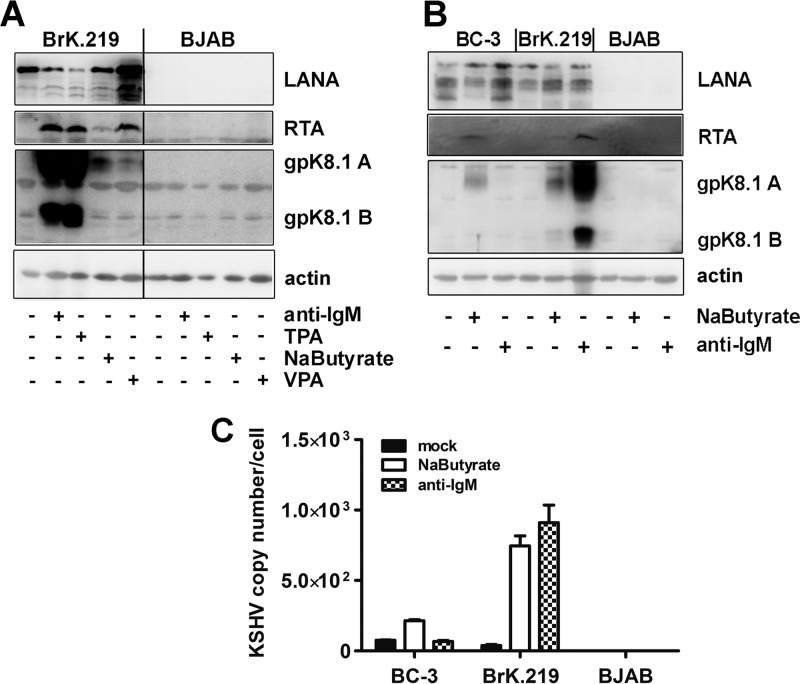
Fig 4.
Comparison of KSHV reactivation induced by antibody against IgM with other chemical stimuli in BrK.219 and in PEL cells. (A) BrK.219 cells and the uninfected parental cell line BJAB were treated with different inducers of the KSHV lytic cycle (2.5 μg of anti-IgM/ml, 50 ng of TPA/ml, 1.25 mM sodium butyrate, 1.25 mM valproic acid [VPA]) for 3 days, and the expression of viral proteins was detected by immunoblotting as described in the legend of Fig. 2. (B) The PEL cell line BC-3, BrK.219 cells, and parental BJAB cells were treated with anti-IgM or sodium butyrate, and the expression of viral proteins was monitored by immunoblotting. (C) KSHV genome copy number in induced BC-3 and BrK.219 cells, as measured by qPCR. The data shown are means of triplicates with the standard deviations.

