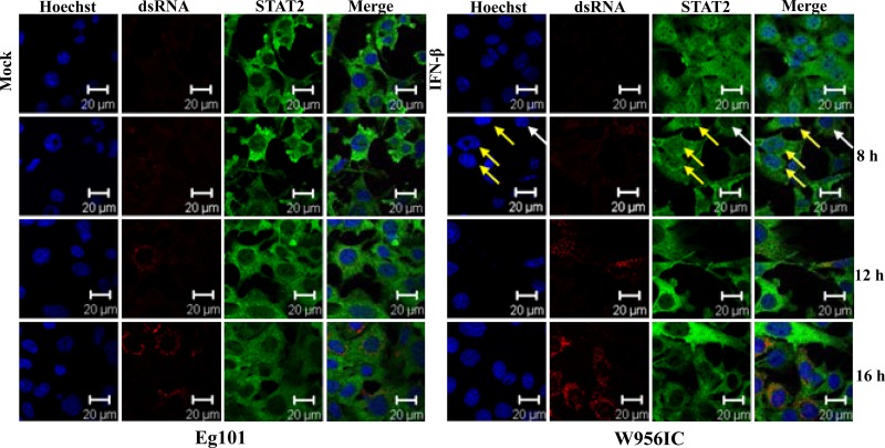
Fig 3.
Analysis of STAT2 nuclear localization. Laser scanning confocal microscopy analyses were performed of STAT2 localization in C3H/He MEFs infected with WNV Eg101 or W956IC at an MOI of 5 for the indicated times or incubated with 1,000 U/ml of IFN-β for 30 min (upper right panel set). Nuclei were stained with Hoechst 33258 dye (blue), STAT2 was detected with anti-STAT2 antibody (green), and infected cells were detected with anti-dsRNA antibody (red). Yellow arrows indicate nuclei that contain STAT2; white arrows indicate a cell with predominantly cytoplasmic STAT2. The results shown are representative of two independent experiments.

