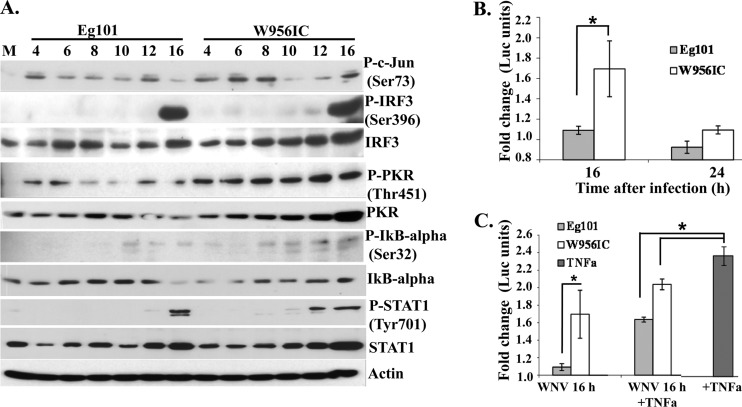
Fig 4.
Activation of transcription factors involved in IFN-β gene induction by WNV Eg101 or W956IC infection in MEFs. (A) C3H/He MEFs were infected with WNV Eg101 or W956IC at an MOI of 1. At the indicated times after infection, cell lysates were prepared, and proteins were separated by SDS-PAGE, transferred, and assayed by Western blotting using specific antibodies. Actin was used as the loading control. The blots are representative of results obtained from three independent experiments. P-, phosphorylated. (B) C3H/He MEFs were cotransfected with an NF-κB luciferase reporter vector (pNF-kB-Luc; Stratagene) and a thymidine kinase (TK) promoter-Renilla vector (pTK-RLuc) using Fugene (Roche) transfection reagent. After 24 h, cells were infected with either WNV Eg101 or W956IC at an MOI of 1. Cells were harvested at the indicated times after infection, and luciferase activity was measured using a dual-luciferase assay kit (Promega). Reporter activity was normalized to Renilla luciferase (Luc) activity in the same sample and is shown as fold change compared to the normalized activity in mock-infected cells. The values shown are averages from three independent experiments. Bars represent ± standard deviations. Asterisks indicate statistically significant differences (*, P < 0.05). (C) At 16 h after infection, mouse TNF-α (20 ng/ml) (Biosource) was added to infected and mock-infected cells for 3 h. The data obtained were analyzed as described for panel B.

