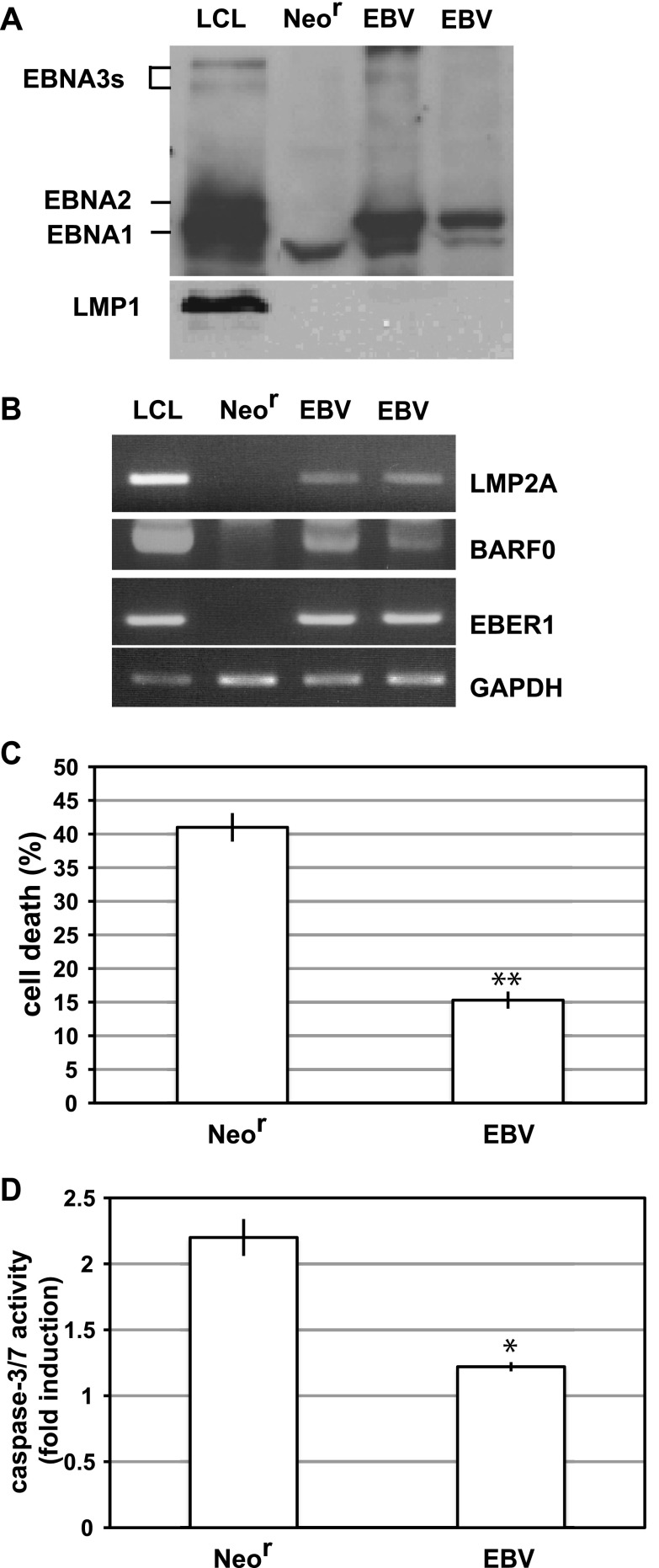
Fig 1.
EBV confers resistance to anoikis in Intestine 407 cells. (A, B) EBV latent gene expression in EBV-positive Intestine 407 cells. (A) Western blot analysis of EBV-positive (EBV) or EBV-negative (Neor) cells. The membrane was probed with EBV-positive human serum for EBNAs and with S12 monoclonal antibody for LMP1. (B) RT-PCR analysis. LCL, a B-lymphoblastoid cell line immortalized with rEBV, was used as a positive control for detection of EBNAs, LMP2A, BARF0, and EBER. (C) Detection of anoikis. EBV-positive (EBV) or EBV-negative (Neor) cells were cultured in suspension for 48 h, and the percentage of dead cells was assessed by Cell Titer-Glo assay. The representative results of 3 separate experiments using 2 independent clones are represented as mean values ± SDs. Statistically significant differences were evaluated by Student's t test (**, P < 0.01). (D) The caspase 3/7 activities of detached cells were measured, and results are represented as the fold induction of the activities of normally cultured cells. The representative results of 3 separate experiments are represented as mean values ± SDs. Statistically significant differences were evaluated by Student's t test (*, P < 0.05).

