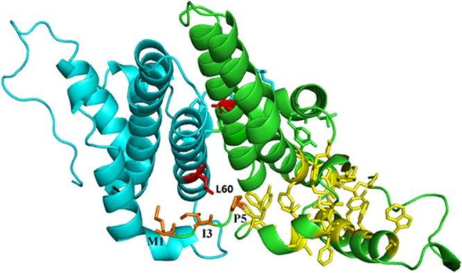
Fig 6.

Structure of the HBV capsid dimer. Hydrophobic residues Met1, Ile3, and Pro5 (orange sticks) and the hydrophobic core (yellow sticks) of one monomer are shown as previously described (40). The two Leu60 residues are shown as red sticks. The structure with PDB accession number 1QGT was used to generate this stereo model using the PyMOL program.
