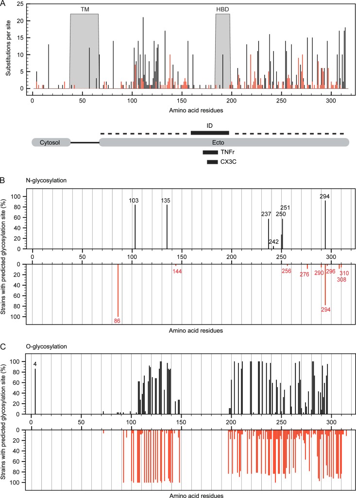
Fig 2.
Substitution hot spots in specific RSV G protein domains and consequences for glycosylation. (A) Schematic representation of the RSV G protein and its specific domains: the transmembrane domain (TM), heparin binding domain (HBD), N-terminal cytosolic domain (Cytosol), C-terminal ectodomain (Ecto), immunogenic domain (ID; aa 159 to 198), CX3C chemokine motif (CX3C; aa 182 to 186), and the region homologous to the fourth subdomain of TNFr (TNFr; aa 171–186). Dashed lines represent mucin-like regions. (B and C) The percentage of strains with predicted sites for N-glycosylation (B) and O-glycosylation (C) within the G protein at a certain amino acid position for both RSV-A (black; 37 strains) and RSV-B (red; 36 strains).

