Fig 2.
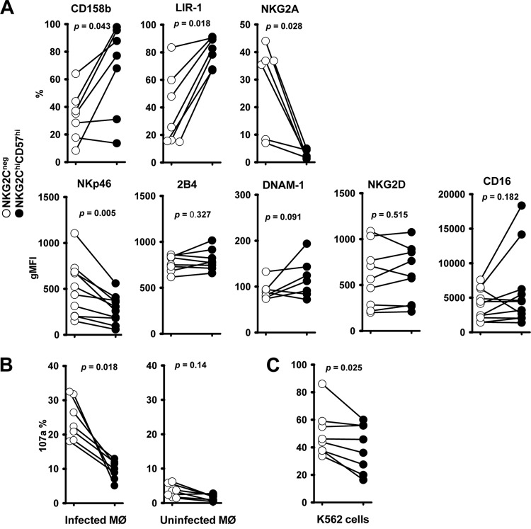
Expression of inhibitory and activating receptors on NKG2Chi CD57hi NK cells (A) and their degranulation to HCMV-infected autologous macrophages (B) and K562 cells (C). (A) Expression of CD158b, LIR-1, NKG2A, NKp46, 2B4, DNAM-1, NKG2D, and CD16 on NKG2Chi CD57hi NK cells and NKG2C-negative NK cells from NKG2Chi CD57hi NK cell-positive donors. The paired circles from one donor are connected by a line. Percentages (CD158b, LIR-1, and NKG2A) or geometric mean fluorescence intensities (gMFI) (NKp46, 2B4, DNAM-1, NKG2D, and CD16) are shown. (B) Thawed PBMCs (1 × 10⁶) were cocultured with TB40/E-infected macrophages (1 × 10⁵) or uninfected macrophages for 48 h. Then, surface expression of CD107a on NK cells was assessed 5 h after the addition of anti-CD107a. (C) Thawed PBMCs (1 × 10⁶) were cultured for 48 h and afterwards cocultured with K562 cells (1 × 10⁵) for 5 h in the presence of anti-CD107a, and then surface CD107a expression was assessed. (A, B, and C) The percentages of positive cells were determined on NKG2Chi CD57hi NK cells and NKG2C-negative NK cells. The nonparametric Wilcoxon signed rank sum test was used to compare NKG2Chi CD57hi NK cells with NKG2C-negative NK cells matched from the same donor.

