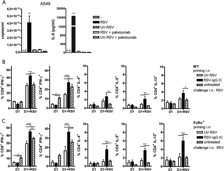
Fig 3.
Intranasal inoculation of IC-RSV efficiently primes RSV-specific CD4+ and CD8+ T-cell responses in both WT and FcRn-deficient mice. (A) Palivizumab neutralizes RSV. RSV (4.7 × 10⁷ PFU) was preincubated for 15 min with 50 μg/ml palivizumab. UV-RSV was used as a negative control for replication. RSV, RSV plus palivizumab, UV-RSV, or UV-RSV plus palivizumab was added to epithelial A549 cells (equivalent of an MOI of 2), and the cells were incubated for 24 h. The results of the RT-qPCR performed on the RSV N gene are shown for one representative experiment of two. Error bars represent the SEM of a duplicate within one experiment. IL-6 production was measured in the 24-h supernatant of A549 cells treated with similar live- and inactivated-RSV preparations. One representative experiment out of 6 is shown. Significance was calculated using a one-way ANOVA. **, P < 0.01. n.d., nondetectable. (B) WT mice and (C) FcRn−/− mice were primed with UV-RSV or IC-RSV at day 0. A third group was untreated. At day 35, all groups were challenged with RSV, and 6 days later, lungs were analyzed for T-cell responses in the presence of uninfected (D1) or RSV-infected D1 (D1+RSV) cells. The percentage of cytokine-producing CD8+ and CD4+ T cells is shown. Error bars represent the SEM of 5 individual mice per group. Results are shown for five mice per group of one of two representative experiments. Significance was calculated using a two-way ANOVA. *, P < 0.05; **, P < 0.01; ***, P < 0.001.

