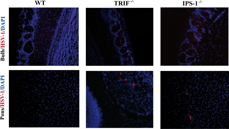
Fig 3.
Representative micrographs illustrating the localization of HSV-1 proteins in different regions of the brains of WT, TRIF−/−, and IPS-1−/− mice (3 mice per group) on day 5 postinfection. Slices (30 μm thick) of brain were processed for immunohistochemistry with a primary polyclonal rabbit anti-HSV-1/2 antibody and a secondary Alexa 594-conjugated goat anti-rabbit antibody (red), followed by staining with DAPI (blue). In WT mice, HSV-1 proteins were restricted to the olfactory bulb and could not be detected in the pons. In TRIF−/− and IPS-1−/− mice, viral antigens were detected in the olfactory bulb and the pons. Magnification, ×20.

