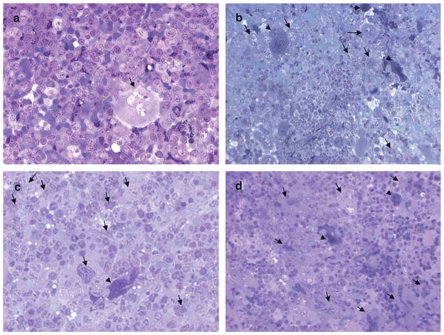
Fig. 1.
MK morphology in the spleens of untreated and TPO-treated WT and Gata1low littermates. Representative semithin sections from the spleen of untreated a,c) and TPO-treated b,d) WT a,b) and Gata1low c,d) mice. Representative megakaryocytes are indicated by arrows. Only one MK is present in the semithin section from WT spleen a). By contrast, the semithin section of TPO-treated WT spleen b) shows nine MKs, six light (whole arrows) and three heavy electron dense (arrowheads). The sections from the spleen of untreated and TPO-treated Gata1low mice are similar c,d). Original magnification: × 40.

