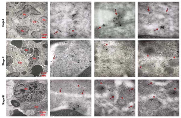
Fig. 10.

Co-localization of VWF and P-selectin in Gata1low MKs at different stages of maturation. Representative sections of spleen from Gata1low mice treated with TPO were double stained for P-selectin (large gold particles) and VWF (small gold particles) and analyzed by immunoelectron microscopy. Red arrows and arrowheads point to structures containing the protein, while the white ones point to the structure itself. In the upper left panel, the nucleus is multilobed, but contains multiple vacuoles while the cytoplasm is immature with abundant RER. Stage II and III Gata1low MKs showed similar ultrastructural features as the cells at the corresponding stage of maturation observed in TPO-treated WT mice (Fig. 8). In the three upper panels, P-selectin and VWF co-localize in the cytoplasm of stage I Gata1low MKs; as in the immature MKs from WT mice (Fig. 8), both proteins can be found in the cytoplasm close to the DMS (whole arrows) and in the primitive α-granules (arrowheads). With progressing maturation, the proteins become increasingly separated, imitating the pattern observed in mature WT MKs. Original magnifications: × 4,400 for the three panels farthest left and × 30,000 for the others.
