Fig. 4.
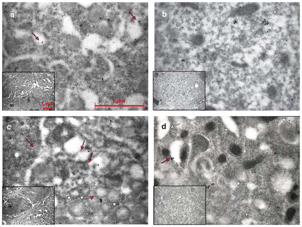
P-selectin distribution in MKs of WT and Gata1low mice before and after TPO treatment. Representative semithin sections of spleen from untreated a,c), and TPO-treated b,d) WT a,b) and Gata1low c,d) mice immunostained for P-selectin and analyzed by immunoelectron microscopy. Representative MKs and the corresponding cytoplasm are shown in the inset and in the panel, respectively. P-selectin staining appears as dots. Red arrows and arrowheads point to structures containing the protein, while the white ones point to the structure as such. As seen in a), P-selectin was found in the α-granule membrane as well as in the cytoplasm in the vicinity of the DMS (whole arrows) in untreated WT mice. After treatment with TPO b), most MKs were immature with only rudimentary DMS and very few granules. a) In these MKs, the major part of P-selectin staining was localized to the cytoplasm. In the immature MKs from the untreated Gata 1 low mice c), P-selectin staining was found both in the cytoplasm and in the scarce α-granules (arrow heads). In the mature forms a), P-selectin was localized primarily in the cytoplasm, often delineating the DMS, but also, atypically, in membranes of vacuolar structures. Original magnifications: × 30,000 for panels; × 4,400 for insets.

