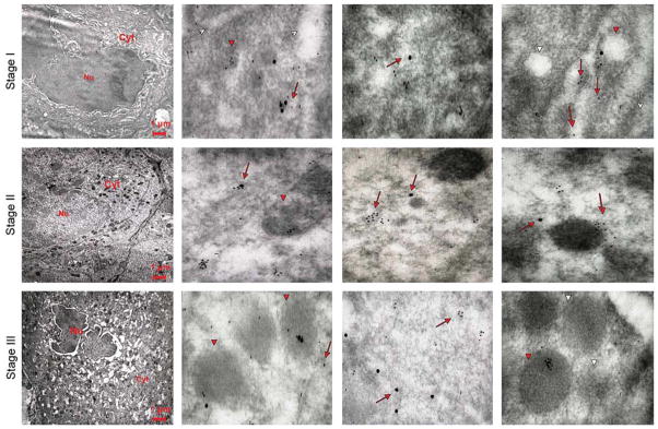
Fig. 8.

Co-localization of VWF and P-selectin in MKs of different stages of maturation observed in WT mice after TPO treatment. Representative sections from WT mice spleens were double stained for P-selectin (large gold particles) and VWF (small gold particles) and analyzed by immunoelectron microscopy. Three representative MKs at different stages of maturation are shown in the left panels, while three representative areas of their cytoplasms are shown in the other panels of each row. Red arrows and arrowheads point to structures containing the proteins, while the white ones point to the structure itself. P-selectin staining appears as isolated dots, and VWF staining shows a punctuate pattern. In a stage I MK, P-selectin and VWF are mostly co-localized, because both proteins are found both in the cytoplasm, often along the DMS (whole arrows), and in the rudimentary α-granule membrane (arrowheads). With maturation, the localizations of the two proteins progressively diverge, and become totally separated in the mature stage III MK (lower panels). Original magnifications: × 4,400 for the three panels on the far left and × 30,000 for the others.
