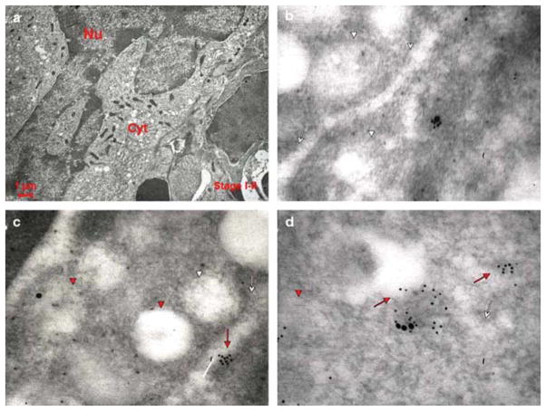
Fig. 9.
Co-localization of VWF and P-selectin in a MK from the spleen of an untreated Gata1low mouse. Representative sections from Gata1low mice spleen were double stained for P-selectin (large gold particles) and VWF (small gold particles). Because the Gata1 mutation blocks maturation (Centurione et al. 2004), only stages I/II MKs were available for analysis. Red arrows and arrowheads point to structures containing the proteins, while the white ones point to the structure itself. In a), cells are characterized by a large nucleus with several nucleoli, with a cytoplasm that contains ribosomes and well developed RER. In b,c,d), P-selectin and VWF co-localize in the cytoplasm in the immature MKs of Gata1low mice. Both proteins were found in proximity to the DMS (whole arrows) and in the vacuoles representing degenerated granules (arrowheads). Original magnifications: a) × 4,400; b,c,d) × 30,000.

