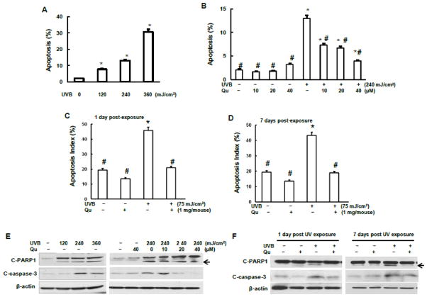
Fig.1.
Effect of quercitrin on apoptosis and apoptotic proteins induced by UVB exposure. A, B, and E, JB6 cells were pretreated with quercitrin (10, 20, and 40 μM) or acetone for 1 h prior UVB exposure. After 24 h, the cells were collected for apoptosis analysis using flow cytometry (A and B) or for immunoblotting assay (E). C and D, 6–8 week old female SKH-1 mice dorsal skin was topically administrated with either aceton (control group) or quercitrin one day before UV exposure. The mice were then exposed to 75 mJ/cm2 of UVB for 3 times per week up to 6 weeks. At 1 day and 7 days, the animals were euthanized and dorsal skin tissues were isolated and subjected for immunofluoresence staining of apoptosis (C and D). The positive cells were counted from a total of 500 cells from 8 random field using Olympus BX51 microscope. The results were expressed as percentage of TUNEL-positive cells (apoptosis index). F, The same as C and D, but mouse dorsal skin tissues were isolated and total protein was extracted for examination of expression levels of cleaved PARP-1 and cleaved caspase-3. * indicates a significant difference compared with control without UVB exposure (p<0.05). # indicates a significant difference compared with 240 mJ/cm2 UVB exposure (p<0.05).

