
Figure 2. KFase fold.
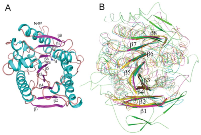
(A) Cartoon presentation of KFase showing an eight-stranded β-sheet surrounded by a number of helices. The catalytic triad is labelled as S (Ser157), H (His276), and D (Asp244) and the β-sheet strands were labelled as β1–β8. The α-helices are shown in cyan, the β-strands in purple and the loops in brown. (B) Superimposition of a carboxylesterase (yellow), an acetyl esterase (orange), a bacterial homologue of the mammalian hormone-sensitive lipase (pink) and human AChE (green) on to KFase (cyan) indicates that all of them have the α/β hydrolase fold signature structure. N-ter, N-terminus.