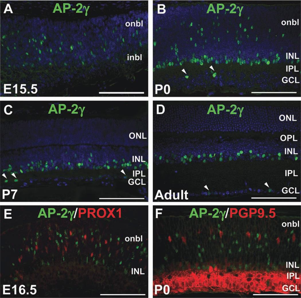
Fig. 3. AP-2γ is not a horizontal cell marker.
A–D: Immunofluorescence using anti-AP-2γ clone 6E4 (green) on horizontal sections of wild-type mouse heads or eyes counterstained with DAPI (4,6-diamino-2-phenylindole (DAPI; blue), at the stages indicated. Arrowheads in B–D indicate AP-2γ-immunoreactive cells in the GCL. E,F: Co-immunostain with anti-AP-2γ (green) and either anti-PROX1 (E) or anti-PGP9.5 (F) antibodies (red) on horizontal sections of wild-type eyes, at the stages indicated. i/onbl, inner/outer neuroblast layer; INL, inner nuclear layer; GCL, ganglion cell layer; IPL, inner plexiform layer; OPL, outer plexiform layer. Scale bars = 100 µm.

